Dongyeol Choi, M.D.
Department of Pathology
Introduction
Traditional pathology has long evolved around optical microscopes and glass slides. However, recent rapid advances in laboratory automation, whole slide imaging (WSI), and artificial intelligence (AI) are fundamentally transforming the paradigm of pathologic diagnosis and laboratory operations as a whole. In particular, in reference laboratories, where large volumes of specimens are continuously submitted or referred from multiple medical institutions, it is necessary to simultaneously manage not only the accuracy of individual tests but also specimen traceability, process standardization, throughput, and turnaround time (TAT). Therefore, the clinical and operational value of digital transformation is even more pronounced in such settings.
In a large-scale reference pathology laboratory, digital transformation does not simply mean a hardware replacement from a “microscope to a monitor.” Rather, it is a process of standardizing the entire workflow, from specimen receipt through tissue processing, embedding, sectioning, staining, high-speed scanning, final interpretation, and result reporting; connecting the flow of physical specimens with digital information; and establishing a foundation on which AI can subsequently be applied. This review examines this process along the continuum of automation–digital pathology–AI and discusses currently available quantitative pathology technologies, the potential of computational pathology to predict molecular information from H&E-stained images, and the practical technical and institutional challenges facing large-scale reference laboratories.
Automation and Digital Pathology: Two Complementary Axes
Many steps in the pathology testing process, including specimen identification, gross examination, tissue processing, embedding, sectioning, staining, and slide sorting and distribution, still involve substantial manual work. As testing volume increases, small inefficiencies and variations at each step accumulate throughout the overall workflow and become a significant operational burden. Therefore, the ultimate goal of laboratory automation is not simply to reduce labor requirements, but rather standardization of the testing process, ensuring specimen traceability, reducing human error, and maintaining stable turnaround times.
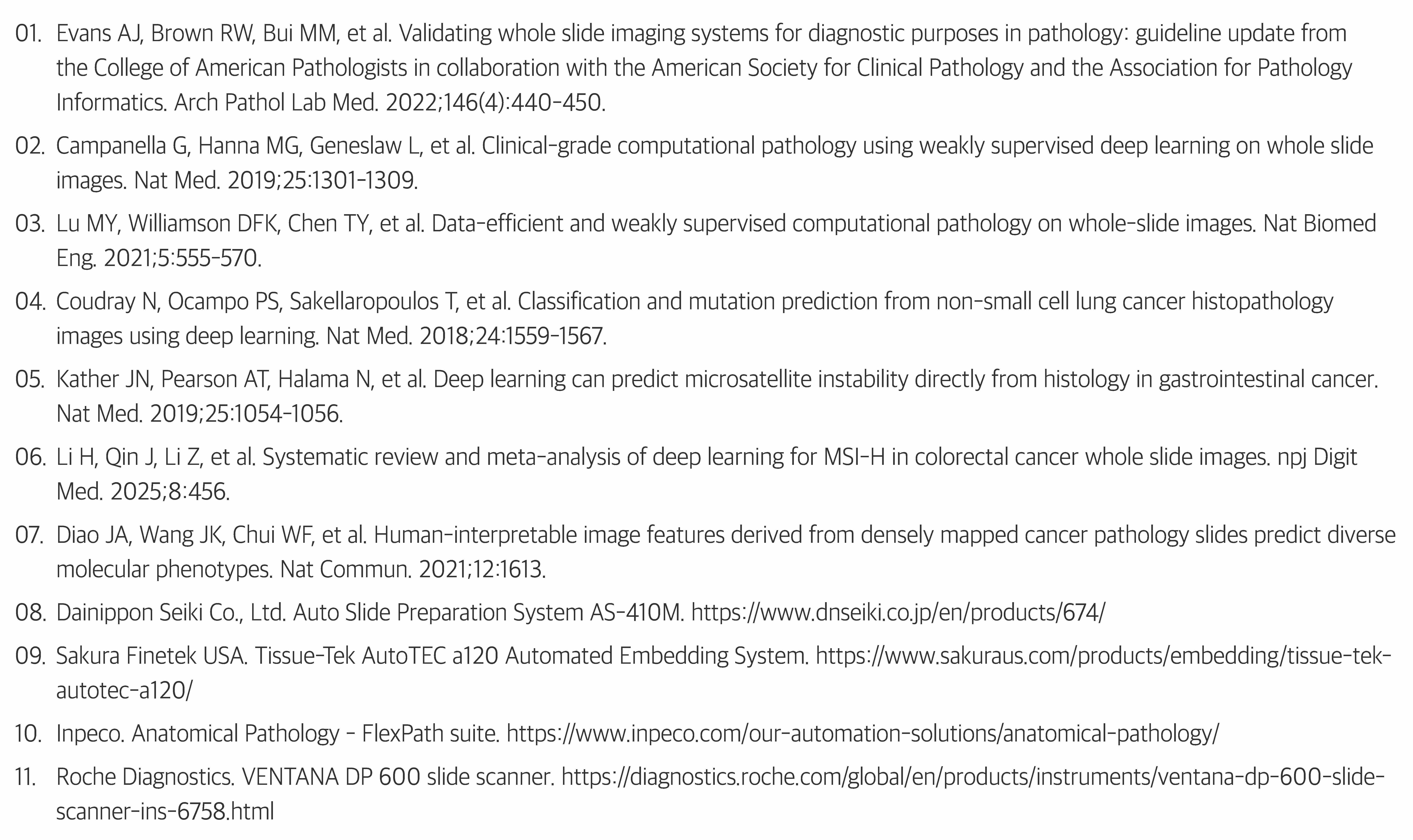
In this context, the Sakura Finetek Tissue-Tek AutoTEC a120 automated embedding system and Dainippon Seiki (DNS) AS-410M automated tissue sectioning system being introduced by our institution are also meaningful. By automating processes such as embedding and sectioning, which are critical determinants of slide quality, operator-dependent variations in tissue orientation, section thickness, and tissue positioning can be reduced, enabling the production of more consistent, high-quality slides.
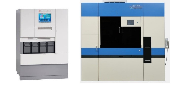
Fig. 1. Sakura Finetek Tissue-Tek AutoTEC a120 automatic tissue embedding system and Dainippon Seiki (DNS) AS-410M automatic microtome system
A barcode-based specimen-cassette-block-slide tracking system and equipment-LIS integration extend this standardization to specimen identification and information flow. In particular, the Trace, Move, and Store systems of the Inpeco FlexPath series integrate the transport, storage, and tracking of formalin-fixed paraffin-embedded (FFPE) blocks into a single workflow, allowing structured information to be retained regarding which processes each specimen has undergone and where it is currently located.
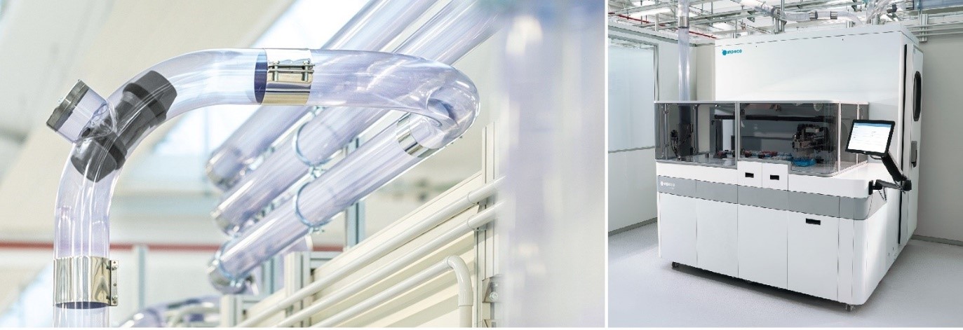
Fig. 2. INPECO FlexPath™ Move and FlexPath™ Store
In this way, while automation standardizes the physical flow of specimens and the pre-analytic phase, digital pathology expands the scope of interpretation and information utilization by converting the slides generated at the end of the process into digital data in the form of whole slide images (WSI). In other words, if automation is the “process of reliably producing standardized slides,” digital pathology can be regarded as the “process of converting those slides into reusable data.”
The use of WSI for diagnosis requires comprehensive validation beyond simply introducing a scanner. The College of American Pathologists (CAP) WSI clinical validation guideline recommends conducting institution-specific validation under conditions similar to the actual clinical environment, using cases appropriate for the intended use and assessing diagnostic concordance with glass slides. In large-scale laboratories, scan throughput, rescan rate, image quality control (QC), case matching, storage capacity, and network bandwidth must also be considered.
Fig. 3. Roche VENTANA® DP 600 slide scanner
The Roche VENTANA® DP 600 slide scanner in use at our institution also serves as a key interface for rapidly converting slides produced through the fully automated process into large-scale WSI data. Once sufficient scanning capacity and stable LIS/IMS integration are secured, not only digital primary diagnosis but also quantitative pathology, automated QC, case triage, and AI for predicting molecular alterations can be incorporated into routine pathology workflows.
The Current State of AI: Quantification and Diagnostic Assistance Rather Than Diagnostic Replacement
The most practical applications of AI in pathology currently lie not in replacing pathologists in making the final diagnosis, but in automating or streamlining repetitive quantitative tasks and supporting diagnostic decision-making. Quantitative pathology, in which tumor regions are detected and cells are classified on WSI to quantify Ki-67 labeling indices and ER/PR, HER2, or PD-L1 expression, is a representative example. Pathologists can review the analysis regions and cell classifications selected by AI, modify them when necessary, and incorporate the results into the final judgment.
The advantages of such quantitative assistance systems extend beyond simple time savings. They can serve as tools for standardization by reducing interobserver/intraobserver variation arising from visual estimation and enabling repeated measurements based on the same criteria. In addition, functions such as tumor detection, metastasis screening, and automatic detection of slide quality abnormalities (slide QC) are highly efficient in reference laboratories where large numbers of cases are received simultaneously. As weakly supervised learning, which trains AI using only slide-level diagnostic information without individually annotating each WSI region, has demonstrated strong classification performance, the possibility of utilizing actual pathology data for AI training without extensive manual annotation is expanding.
However, when AI is deployed as a standalone application, it may instead complicate the workflow. If a pathologist must check clinical information in the LIS, open the WSI in another viewer, review AI results in yet another program, and then return to the LIS, the process may be digitally enabled but operationally less efficient. Therefore, an ideal operating system is one in which AI should be embedded directly within the IMS or primary viewer, automatically invoked for appropriate cases for the necessary cases, and its results are stored together with diagnostic information.
From Morphology to Molecular Information: The New Value of H&E Images
The more fundamental change that AI can bring to pathology does not stop at rapidly performing quantitative measurements previously conducted by humans. H&E slides contain the morphology of tumor cells, nuclear structure, cellular arrangement, stroma, the distribution of immune cells, and the spatial relationships within the tumor microenvironment. Because these morphologic phenotypes reflect underlying genetic alterations, gene expression patterns, and cellular biology, digital images may contain molecular information that is difficult for the human eye to recognize clearly.
In 2018, Coudray et al. reported that a deep learning model using H&E WSI of lung cancer could not only distinguish lung adenocarcinoma from squamous cell carcinoma but also predict STK11, EGFR, FAT1, SETBP1, KRAS, and TP53 mutations in lung adenocarcinoma with AUCs (area under the receiver operating characteristic curve) ranging from 0.733 to 0.856. AUC is a metric indicating how well a model distinguishes cases with a particular abnormality from those without it; 0.5 represents performance no better than chance, whereas 1.0 represents perfect discrimination. These findings suggested a new paradigm in which, in addition to the traditional workflow of “performing molecular testing after morphological diagnosis,” the possibility of a molecular abnormality can first be estimated from H&E images, thereby allowing cases to be triaged for confirmatory molecular testing.
In gastrointestinal tumors, microsatellite instability (MSI) prediction is a representative example. Kather et al. demonstrated that microsatellite instability (MSI) could be directly predicted using deep learning from H&E images of colorectal and gastric cancers, and subsequent multicenter studies and external validations have followed. According to a 2025 meta-analysis of WSI-based MSI-H prediction in colorectal cancer (19 studies, 33,383 specimens), pooled sensitivity of 0.93 and pooled specificity of 0.71 were reported in external validation. The high sensitivity and relatively low specificity suggest that the current role of this technology is more suitable as a screening tool for triaging high-risk cases rather than replacing molecular testing.
The scope of research is expanding beyond the prediction of individual single-gene mutations to molecular phenotypes and gene expression. Diao et al. demonstrated that various molecular characteristics, including immune checkpoint protein expression and homologous recombination deficiency, could be predicted by quantifying pathologist-interpretable morphologic features from WSI of five cancer types. More recently, end-to-end workflows for directly predicting biomarkers from WSI have been proposed as standardized research protocols, indicating that computational pathology has developed into an independent research field.
The key point of this change is that pathology images are no longer merely “pictures viewed by humans.” Once a slide becomes a WSI, the image becomes computable data, and AI can extract features from that data that humans have not explicitly defined. Therefore, the question of the future may be less about “whether AI can replace pathologists” and more about “how much more clinically useful information can be obtained from H&E that could not previously be obtained

Fig. 4. From AI-based morphological feature extraction from H&E-stained slide images to molecular information prediction and subsequent clinical application
The Unique Data Advantage of Large-Scale Reference Laboratories
The key resource for computational pathology is not algorithms alone, but large-scale, high-quality, clinically curated multimodal data. Simply possessing a large number of H&E WSI is not sufficient; each image must be accurately linked to results from final pathologic diagnosis, immunohistochemistry (IHC), FISH, PCR, NGS, and other tests. Large-scale reference laboratories have unique potential because large volumes of specimens are received from diverse medical institutions and multiple types of pathology and molecular tests are performed within a single institution.
In particular, if H&E WSI from the same patient are structurally linked to biomarker and genomic analysis data, current test results can serve as high-quality ground-truth data for the development of next-generation AI models in the future. If process metadata, including specimen identifiers, instrument parameters, staining conditions, and reagent lot numbers collected in real time by laboratory automation systems, are also linked, this can help trace differences in image quality and the causes of AI errors. This represents a strength distinct from that of university hospitals. University hospitals are advantageous for securing detailed treatment information and long-term outcome data, whereas large-scale reference laboratories can accumulate very large-scale real-world pathology data across broad ranges of institutions and patient populations. If data linkage with medical institutions becomes possible in the future, these two types of data could be combined to create even greater value.
Therefore, a more important question than the testing volume itself is not “How many slides are held?” but “How many high-quality WSI are held with reliable diagnostic, immunohistochemical, and molecular pathology results?” The linkage structure must be designed together with the current digital pathology implementation process in order for these data to acquire value as a data asset.
Challenges to Be Addressed by Large-Scale reference laboratories
1. Standardization of Slide Quality
WSI quality and AI performance begin upstream, with the quality and consistency of the entire pathology workflow. Uneven tissue section thickness, variation in staining intensity, air bubbles and wrinkles, paraffin contamination, and inadequate mounting can affect not only scanning success rates but also AI performance. Humans can adapt to a certain degree of variation, whereas AI may be highly vulnerable to differences between training data and actual input data, namely domain shift. Therefore, the effects of introducing embedding and sectioning automation equipment such as the AutoTEC a120 and AS-410M need to be continuously evaluated not only in terms of throughput but also in terms of section quality uniformity, rework rate, scanning success rate, and other indicators. In the AI era, standardization of tissue processing and slide preparation and management of quality indicators are becoming even more important.
2. Workflow Design to Prevent Scanning Bottlenecks
A fully digital workflow introduces new steps after slide preparation, including scanning, image QC, case-to-image matching / slide-to-case matching, and large-volume file transfer. Even when high-capacity scanners are introduced, relying solely on nominal processing speed (slides/hour) may substantially overestimate real-world operational capacity. Actual processing time, including focus errors, barcode recognition failures, tissue detection failure, rescanning, processing of large slides, and slide loading/unloading, must be measured. In large-scale environments, even 1–2% of exceptions can generate dozens of manual tasks every day. Therefore, the operational performance of scanners, including the Roche VENTANA DP series, needs to be managed using indicators such as first-scan success rate, rescan rate, manual intervention rate, and the time required until an image is actually available for diagnostic interpretation after scanning.
3. Integration of Automation–Scanner–LIS/IMS
If completed slides in an automated laboratory are manually sorted again and loaded into the scanner by personnel, and scanned images are manually linked to cases, a critical gap remains between physical automation and the digital workflow. The identifiers for specimens, cassettes, blocks, and slides obtained from an intelligent logistics tracking system such as Inpeco needs to be seamlessly linked as WSI metadata, and scanned images need to be automatically linked to the corresponding case and the pathologist’s worklist. LIS, IMS/viewer, and AI inference engines should also be connected within a single diagnostic workflow rather than being operated as siloed systems.
4. Machine-Readable Integration of WSI and Multimodal Test Results
Even if large testing institutions generate large amounts of data every day, it is difficult to use the data for AI training if the results are scattered across free-text reports, PDFs, and separate databases. At a minimum, case identification (case ID), final pathological diagnosis, cancer classification, biomarker testing methods, detailed genetic variant information, and WSI files should be interlinked in a machine-readable structure. Data structuring is not an additional task for future research; it is a core infrastructure that must be designed together during the digital pathology implementation stage.
5. In-House Clinical Validation and Continuous Quality Monitoring
There is no guarantee that an AI demonstrating an excellent AUC in a research paper will achieve the same performance in an actual laboratory. Differences in tissue processing, staining reagents and concentrations, scanner sensor characteristics, image compression algorithms, and the disease composition of the incoming patient population can affect performance. Therefore, AI implementation should be approached similarly to the introduction of a new testing method, with the intended use defined and institution-specific validation performed. In addition, a continuous monitoring system is required to monitor changes in performance when staining protocols or scanners are changed. The relatively lower specificity observed in external validation in the 2025 MSI-H meta-analysis clearly demonstrates that generalizability and standardization remain important challenges.
Conclusion: A Leap from Automation to Precision Pathology
The future of large-scale reference pathology laboratories does not lie in pursuing automation, digital pathology, and AI as separate projects. These three technologies should be viewed as a single continuum consisting of “the stage of standardizing and tracking specimens – the stage of converting slides into digital data – and the stage of extracting new information from that data.”

Fig. 5. Digital Transformation of Large-Scale Reference Pathology Laboratories: From Automation to Computational Biomarkers
The most realistic current applications of AI are supportive functions such as quantitative pathology, tumor detection, slide quality control, and triage of high-risk cases. At the next stage, H&E images may be used to predict the likelihood of MSI or specific genetic abnormalities and thereby select cases for additional testing. Furthermore, as multimodal AI that simultaneously analyzes H&E morphology, digital immunopathology (IHC), large-scale genomic data, and patient clinical information advances, it may expand into a tool for precision oncology that predicts prognosis and treatment response.
Realizing this vision does not require waiting for fully mature AI systems. Standardized slides must be produced reliably through automated embedding and sectioning equipment; the entire transport process of blocks and slides must be tracked in real time; sufficient scanning capacity and rigorous QC systems must be established; and LIS and IMS must be connected so that the generated WSI can be accumulated in a structured form accurately linked to pathological and molecular results. When each automation investment currently being pursued is connected within this continuum, its impact can extend beyond productivity improvement to the creation of the data infrastructure required for future AI development and training. Today’s automation creates the foundation for digital pathology, and today’s digital pathology creates tomorrow’s AI training data.
From this perspective, digital transformation of the pathology laboratory is not simply the process of creating a “laboratory without microscopes.” It represents a transition toward a new pathology workflow that converts pathology slides into computable digital data, identifies hidden information within that data, and returns it to patient diagnosis and treatment. If the testing scale and diverse testing portfolio of large -scale reference laboratories are systematically connected, their role can extend beyond operational efficiency to becoming a leading hub for the discovery of next-generation computational biomarkers and the realization of precision pathology.
References